Primary care CME: Understanding the importance of a 12-lead ECG
One of the aspects of Primary care CME and emergency care CME is to understand the importance of a 12 lead electrocardiogram. It is critical to understand how a 12 lead ECG can help in primary and emergency care. That will enable you to pay more attention to the readings of ECGs to detect and determine your patients’ vital health signs that could otherwise go unnoticed. Here’s more on the importance of a 12-lead ECG in primary and emergency care.
12 lead ECG – what it is and how to use it in primary and emergency care
An electrocardiogram records the heart’s electrical activity from various angles to identify and locate multiple heart conditions. Electrodes placed on the chest painlessly record the heart’s electrical impulses.
ECG leads are graphical representations of the electrical activity of the heart.
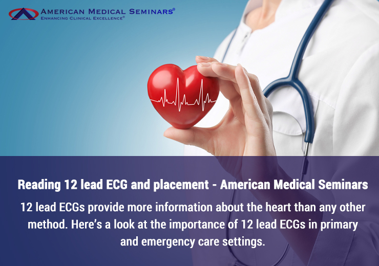
In the case of a 12-lead ECG, 10 physical electrodes are placed on the chest, ankles, and wrists. The electrodes are connected to a monitor that records the electrical activity in the heart. The ECG generates 12 perspectives or leads (from the 10 electrodes). Those 12 leads provide more information about heart conditions than any other procedure.
A 12 lead ECG can screen patients for any heart condition or heart attack in primary care and emergency care settings to get appropriate and immediate medical intervention. In some cases, a 12-lead ECG is the only marker for the presence of a heart condition.
When is a 12 lead ECG required?
Healthcare professionals perform ECGs in primary and emergency care settings for various reasons:
1. Check the electrical activity of the heart.
2. Find the reason for any unexplained chest pain. The reasons could range from a heart attack to angina or even pericarditis.
3. Find the cause for symptoms of heart disease. The patient’s symptoms include shortness of breath, fainting, dizziness, palpitations, or rapid heartbeats.
4. To detect any hypertrophy of the walls of the heart chambers.
5. To ensure medicines are working correctly or might be causing side effects that might affect the heart.
6. To check and ensure mechanical devices like pacemakers are working correctly to ensure a normal heartbeat.
7. To determine the heart’s health when a patient has underlying conditions such as diabetes, high cholesterol, a family history of heart disease, or high blood pressure.
Placement of electrodes
Understanding the placement of electrodes is a part of all excellent primary care CME or emergency care CME. Here is a quick look at the placement of each electrode.
Chest electrodes and their placement
Number of electrodes: 6 electrodes, V1 to V6
Placement:
V1: To be placed along with the 4th intercostal space (at the right sternal edge).
V2: To be placed at the 4th intercostal space (at the left sternal edge).
V3: To be placed midway between V2 and V4.
V4: To be placed at the 5th intercostal space (in the midclavicular line)
V5: To be placed at the left anterior axillary line (at the same horizontal level as the V4)
V6: To be placed along the left mid-axillary line (at the same horizontal level as V4 and V5)
Limb electrodes and their placements
Number of limb electrodes: Four electrodes – right arm (RA), left arm (LA), Left leg (LL), right leg (RL)
Placement
Right arm (RA): Placed anywhere between the shoulder and elbow of the right arm.
Left arm (LA): Placed anywhere between the shoulder and elbow of the left arm.
Left leg (LL): Placed anywhere below the left torso and above the left ankle.
Right leg (RL): Placed anywhere below the right torso and above the right ankle.
Understanding the representation of the leads
As a primary care or emergency care PA, it is critical to understand the various anatomical territories of the heart and which lead represents them. Primary care CME and emergency care CME cover this in detail. Here is a quick view of the leads and which view of the heart they provide.
The Chest leads
V1 and V2 provide septal views of the heart.
V3 and V4 provide anterior views of the heart.
V5 and V6 provide lateral views of the heart.
The other leads
Lead I, aVL, and aVR provide lateral views of the heart.
Lead II, III, and aVF provide inferior views of the heart.
As a healthcare professional working in the emergency or primary care wings, it is mandatory to understand how to conduct a 12-lead ECG and read the results correctly. While a 12-lead ECG is a blessing in primary and emergency care settings, misreading an ECG can lead to wrong diagnoses and clinical decisions. For this reason, ECGs are a part of all primary care CME and emergency care CME courses.
American Medical Seminars has been a leading provider of concise, to-the-point, clinically-relevant CME courses for over three decades. All the courses are in downloadable audio and video formats. Courses can be taken anytime, anywhere, and at one’s own pace.